Corticobasal ® Implants: Immediate Loading Following Mandibulectomy
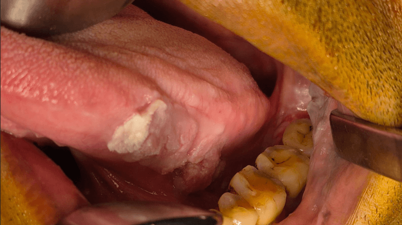
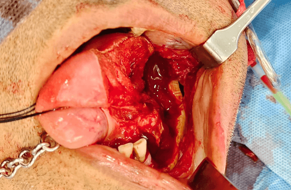
Abstract Background: Resections of the mandible, whether due to benign aggressive lesions or malignant neoplasms, drastically alter the biomechanical and anatomical landscape of the stomatognathic system, leading to profound deficits in mastication, speech, and aesthetic appearance. The rehabilitation of patients following marginal or segmental mandibulectomy represents one of the most formidable challenges in maxillofacial prosthodontics, particularly when surgical intervention is compounded by adjuvant radiotherapy. Traditional reconstructive paradigms often rely on complex vascularized grafting followed by delayed placement of conventional crestal implants, a process that prolongs patient morbidity and frequently contraindicates immediate functional restoration. This paper outlines a comprehensive clinical and methodological framework for utilizing corticobasal ® implants to achieve immediate functional loading in post-mandibulectomy patients. Technology of Strategic implants® offer an alternative to conventional endosseous implants, with potential advantages in compromised bone and irradiated fields due to their bicortical anchorage, engaging the highly mineralized, vascular-independent basal bone and immediate loading capability. Keywords: corticobasal implants, Strategic Implants, immediate loading, marginal mandibulectomy, oral cancer reconstruction, dental rehabilitation, radiotherapy, cortical anchorage 1. Introduction Surgical management of mandibular pathologies — ranging from benign aggressive odontogenic tumors to malignant squamous cell carcinomas — frequently necessitates marginal or segmental mandibulectomy. Marginal mandibulectomy preserves the inferior mandibular border while sacrificing the alveolar ridge; segmental mandibulectomy involves full-thickness resection, disrupting arch continuity. Both procedures compromise masticatory function, speech, lower lip support, and aesthetics, imposing significant burden during an already demanding period of recovery. The four patients in this series reflect the breadth of pathologies encountered in clinical practice: verrucous carcinoma, squamous cell carcinoma of the floor of mouth, squamous cell carcinoma of the lower alveolus, and unicystic ameloblastoma. Despite differing diagnoses, all shared a common rehabilitative challenge and a common anatomical resource — the preserved inferior cortex of the mandible. Conventional implant rehabilitation in resected mandibles depends on adequate alveolar bone volume and cancellous vascularity, both frequently absent in post-resection patients. Where adjuvant radiotherapy has been administered, radiation-induced osseous changes further elevate the risk of implant failure. Traditional protocols also demand prolonged multi-stage timelines, leaving patients edentulous and functionally compromised for months. Corticobasal implants ® address these limitations by engaging the dense basal cortex rather than the deficient alveolar crest, providing primary stability sufficient for immediate loading within 48–72 hours. This approach was applied across all four cases — in three as primary post-resection rehabilitation, and in one as salvage following failure of conventional implants in a fibula-reconstructed neomandible. This series documents the clinical presentations, implant protocols, prosthetic outcomes, and follow-up findings of these four cases, discussing the anatomical and biological rationale for the technology of strategic implants ® in the post-mandibulectomy patient. 2. Case Descriptions Case 1: Verrucous Carcinoma with Marginal Mandibulectomy 2.1 Presentation and Diagnosis A 46-year-old male presented with a non-healing ulcer on the left lateral border of the tongue (Fig.1) of approximately 3 months' duration. Incisional biopsy confirmed verrucous carcinoma and PET-CT demonstrated a small ulcerative lesion in the mid-third of the left lateral border of the tongue with no cervical lymph node uptake. The patient underwent left adequate glossectomy and marginal mandibulectomy of the left mandibular body (Fig. 2) under general anaesthesia, along with selective neck dissection of levels I–IV on the left side and free radial forearm flap (FRAFF) reconstruction. Resection dimensions measured 4.2 × 1.8 cm with histologically confirmed clear margins on frozen section. Histological examination confirmed early invasive squamous cell carcinoma with keratinising high-grade dysplasia. The patient returned after 3 months requesting dental rehabilitation for improved function. The OPG (Fig. 3) showed intact lower border of mandible with all teeth missing in the third quadrant except 31.
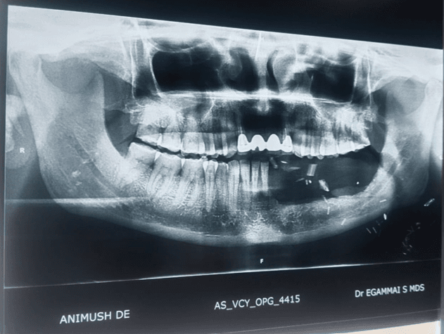
2.2 Implant Procedure The flap was debulked along with sulcoplasty, and a CO₂ soft tissue laser was used for hair removal. Four BCS implants (3.6 mm diameter; lengths 14 mm, 17 mm, 12 mm, 12 mm) were placed (Fig. 4) engaging the inferior cortex of the residual mandibular base. The implants were bent to achieve parallelism between abutments. An intraoral scan was performed after placing scan bodies, and putty was used as a spacer (Fig.5) until the definitive appointment.
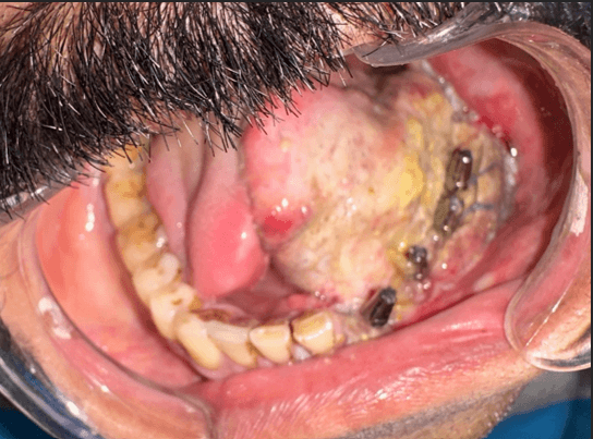
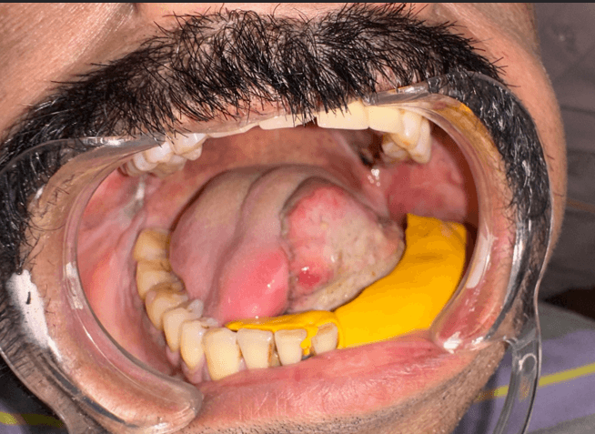
2.3 Prosthetic Rehabilitation and Follow-up A fixed provisional HIPC bridge (Fig. 6) with hygiene pontic was delivered at 48 hours postoperatively. Healing was uneventful. Occlusion was restored with heavy contacts on the right side and minimal contact on the left during mandibular closure. The patient reported high satisfaction with aesthetic and functional outcomes. At 6-month follow-up, the patient was comfortable.
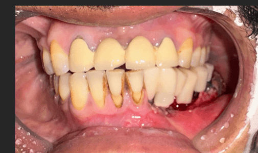
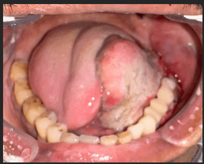
Case 2: Squamous Cell Carcinoma of the Floor of Mouth with Adjuvant Radiotherapy 2.4 Presentation and Diagnosis A 37-year-old male presented in August 2020 with a non-healing lesion over the right floor of mouth of 3 months' duration. PET-CT demonstrated an irregularly enhancing ulcerative mass measuring 3.2 × 2.4 cm involving the floor of mouth, and biopsy confirmed well-differentiated squamous cell carcinoma. He underwent wide local excision of the floor of mouth lesion, marginal mandibulectomy, partial glossectomy, bilateral selective neck dissection, free radial forearm flap reconstruction, and tracheostomy in September 2020. Four BCS implants were placed in native bone at the time of surgery with a provisional prosthesis (Fig. 8 & 9) to maintain lower lip support. He subsequently underwent adjuvant radiotherapy in November 2020.
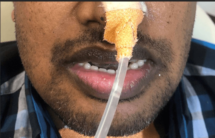
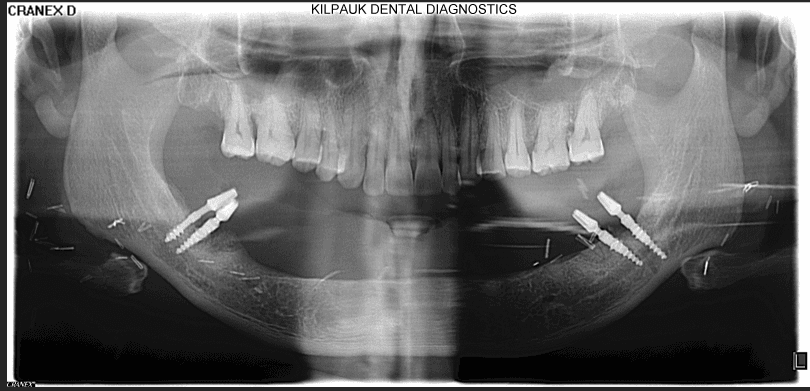
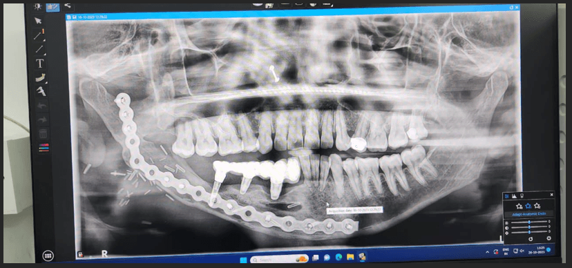
2.5 Implant Procedure and Prosthetic Rehabilitation The provisional denture proved non-retentive and non-functional, prompting the patient to return in 2023 for a definitive solution (Fig. 10). Six BCS implants were placed in the lower jaw through the flap, and impressions were made using impression caps (Fig. 11). A metal framework try-in (Fig. 12) was performed to verify fit, following which a metal-composite hybrid prosthesis (Fig. 13) was cemented. Occlusion was adjusted and dietary instructions provided.
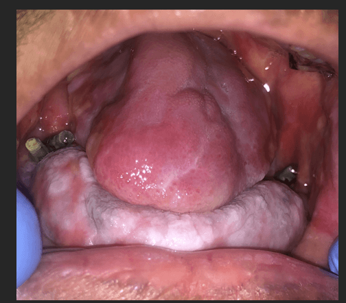
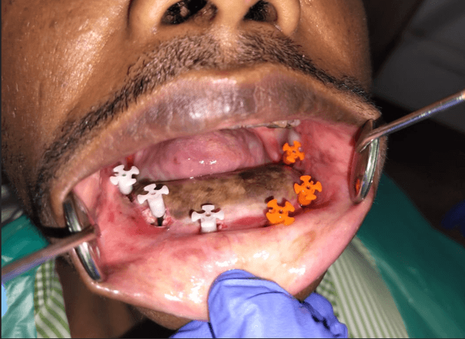
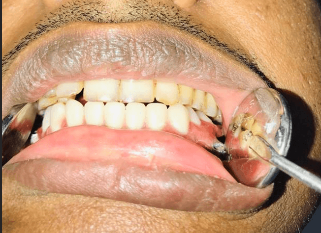
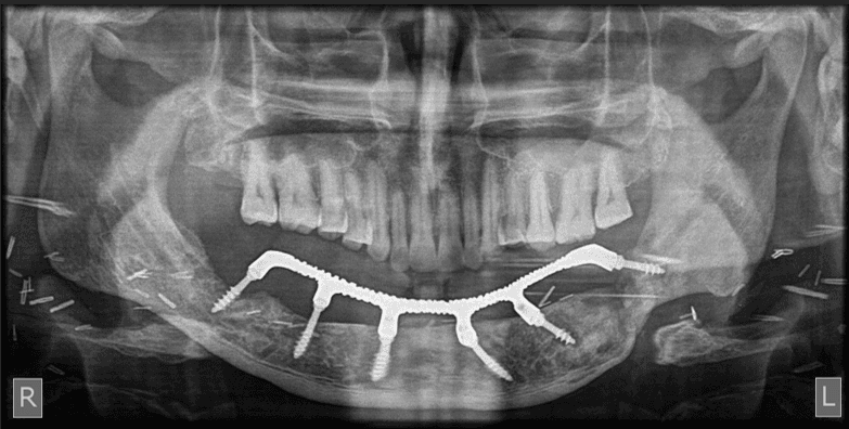
2.6 Follow-up The patient was followed up through December 2025, with implants remaining clinically stable throughout the follow-up period (Fig.14 & 15). No osteoradionecrosis or implant failure was recorded.
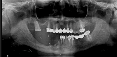
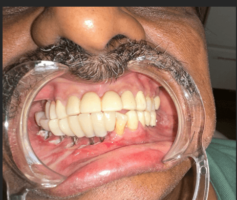
Case 3: Squamous Cell Carcinoma of the Lower Alveolus 2.7 Presentation and Diagnosis A 67-year-old male presented with an ulcerative lesion in the right lower alveolus of 6 months' duration. The ulcer had been precipitated by sharp margins of an existing dental restoration. He underwent right marginal mandibulectomy (Fig.16) with right neck dissection and FAMM flap reconstruction in 2023. Histopathological examination confirmed moderately differentiated squamous cell carcinoma.
2.8 Implant Procedure and Prosthetic Rehabilitation The patient returned in June 2024 for prosthetic rehabilitation. The left occlusal plane was established before proceeding with the prosthetic rehabilitation on right side (Fig.17) Six BCS implants were placed (Fig. 18), impressions were taken, and a G-CAM prosthesis was delivered following occlusal adjustment.
2.9 Follow-up The patient attended follow-up appointments at 3 months, 6 months, and 1 year. Peri-implant mucositis was noted at the 3-month review (Fig. 19), attributed to a thick flap predisposing to bacterial colonisation around the implant collar. This resolved completely with non-surgical management and enhanced oral hygiene instruction. No further complications were recorded.
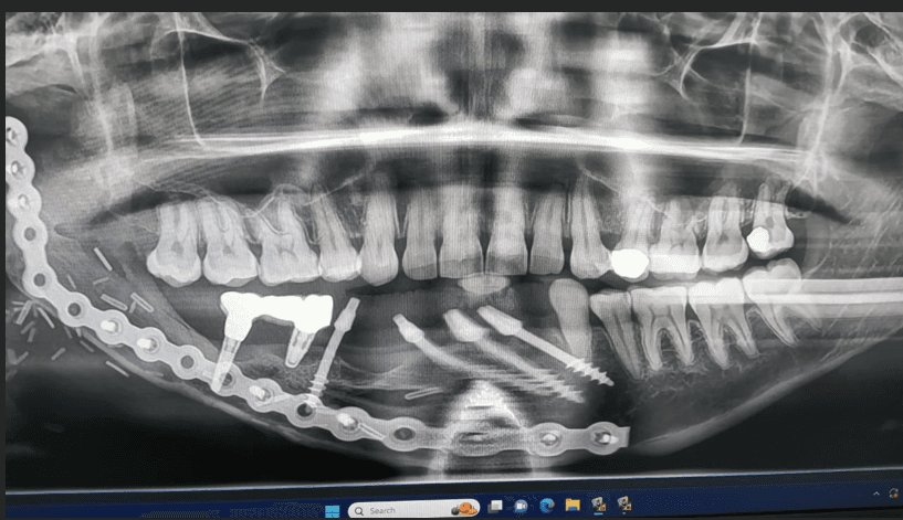
Case 4: Unicystic Ameloblastoma — Salvage Following Failed Conventional Implants in Fibula Neomandible 2.10 Presentation and Diagnosis A 42-year-old female presented with a slowly progressive, painless swelling on the right side of the lower jaw of 8 months' duration, with associated mild facial asymmetry. CBCT confirmed cortical expansion with focal perforation, involvement of the mandibular canal, and lesion extent from the right first premolar to the angle of the mandible. Histopathological examination confirmed unicystic ameloblastoma, mural subtype, with follicular pattern demonstrating peripheral palisading of columnar cells with reverse polarity and stellate reticulum-like cells. Segmental mandibulectomy was performed removing the right mandibular body from the first premolar to the angle. The inferior alveolar nerve was sacrificed due to involvement; frozen section margins confirmed tumor-free bone bilaterally. A reconstruction plate was secured to the residual segments and a double-barrel fibula free flap was employed, with the skin paddle used for floor-of-mouth mucosal reconstruction. At 6 months post-reconstruction, three conventional endosseous implants were placed in the fibula neomandible and submerged for 3 months. Following second-stage surgery and prosthetic loading with a metal-ceramic prosthesis, progressive periimplant bone loss was observed (Fig.20). At 8 months, prosthesis mobility prompted sectioning of the bridge and removal of one failed implant.
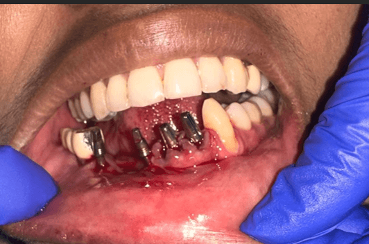
2.11 Corticobasal Implant Procedure and Prosthetic Rehabilitation Following a 3-month healing period after explantation, four corticobasal implants (Fig. 21) were placed with bicortical fixation engaging both the superior and inferior cortical plates of the fibula segment and the native mandible in the anterior region. The three lower incisors were extracted (Fig.22) to facilitate anterior implant placement. Immediate loading was performed at 72 hours with a transitional G-CAM splinted prosthesis to distribute occlusal forces and minimize micromovement during osseointegration.
3. Discussion The four cases presented illustrate that corticobasal implants ® with immediate functional loading can be successfully integrated into the rehabilitative pathway following mandibulectomy across a range of benign and malignant pathologies. The unifying anatomical prerequisite — preservation of the inferior mandibular cortex — enabled bicortical anchorage in all cases regardless of aetiology or resection extent. A recurring theme across cases is the relationship between resection geometry and implant positioning. In cases involving the anterior mandible (Cases 3 and 4), the symphyseal and parasymphyseal cortex provided dense bicortical bone of adequate height despite the greater aesthetic demands of the anterior arch. In the posterior cases (Cases 1 and 2), the inferior cortex of the body and angle offered a reliable fixation zone even where lateral expansion or thinning was present. Case 2 merits particular discussion with regard to the irradiated field. Prosthetic rehabilitation in this patient served a dual purpose: restoring masticatory function and supporting nutritional and psychosocial wellbeing during oncological recovery. No osteoradionecrosis or implant failure was recorded within the follow-up period, consistent with observations that basal cortical bone — with its richer blood supply and lower metabolic turnover compared to alveolar bone — may be comparatively less susceptible to radiation-induced vascular compromise. Nevertheless, given the established risk of osteoradionecrosis in irradiated mandibles, long-term vigilance and extended follow-up remain mandatory in such cases. Case 3 was the only patient to develop a complication — peri-implant mucositis at 3 months — which resolved fully with non-surgical treatment. The thick reconstructive flap in this patient predisposed to bacterial colonisation around the implant collar, underscoring the importance of enhanced early oral hygiene instruction and more frequent review intervals in patients with bulky soft tissue reconstruction. Case 4 illustrates the utility of corticobasal implants as a salvage strategy following conventional implant failure in fibula-reconstructed bone. The reduced bone density inherent to the double-barrel fibula configuration, combined with altered periosteal vascularity, likely contributed to the failure of conventional endosseous implants. Bicortical anchorage in both the fibula cortex and native anterior mandible provided the primary stability required for immediate loading, ultimately achieving the functional and aesthetic rehabilitation that conventional implants had failed to deliver. This series has important limitations. The small number of cases, retrospective design, absence of a comparison group, and relatively short follow-up period limit the generalisability of findings. Larger prospective studies with five or more years of follow-up are needed to assess long-term bone stability — particularly in irradiated cases — and to establish formal indications and protocols for corticobasal implantology in post-mandibulectomy rehabilitation. 4. Conclusion This case series demonstrates that corticobasal implants with immediate functional loading represent a clinically viable and reproducible rehabilitative strategy following marginal and segmental mandibulectomy. By engaging the preserved basal cortex rather than the deficient alveolar crest, this approach circumvents the principal anatomical limitations of conventional implants in post-resection bone. Immediate loading within 48–72 hours meaningfully reduces the period of functional edentulism, a relevant advantage in oncological patients facing concurrent nutritional and psychosocial challenges. The successful salvage of a failed conventional implant case in a fibula-reconstructed neomandible further expands the potential clinical application of this technique. Prospective studies with longer follow-up are warranted to confirm these outcomes at scale.
Leave a Reply
Your email address will not be published. Required fields are marked *
Comments (0)
No comments yet. Be the first to share your thoughts!